What is Melanoma?
Melanoma is a type of skin cancer that originates in the melanocytes of the top layer of skin or the epidermis.
There are three types of cells located in the top layer of skin, which include squamous cells, basal cells, and melanocytes. The melanocytes are often darkly colored due to the melanin pigment in the skin, though there are cases in which the melanocytes can be pinker or whiter in appearance. Most often, melanoma is darker in color due to its exposure to sunlight and ultraviolet (UV) rays. Melanoma occurs when there has been irreparable damage to melanocytes’ DNA, causing the melanocytes to divide and multiply uncontrollably. The most critical factor in learning about melanoma is its ability to spread throughout the body. Melanoma can spread through the bloodstream and travel to distant organs, something that the other forms of skin cancers (basal and squamous cell carcinomas) cannot do. A dermatologist, or a medical doctor who specializes in skin disorders, is usually the healthcare professional who makes the melanoma diagnosis, but at times, any medical office that can send specimen(s) to a dermatopathologist can make the diagnosis.
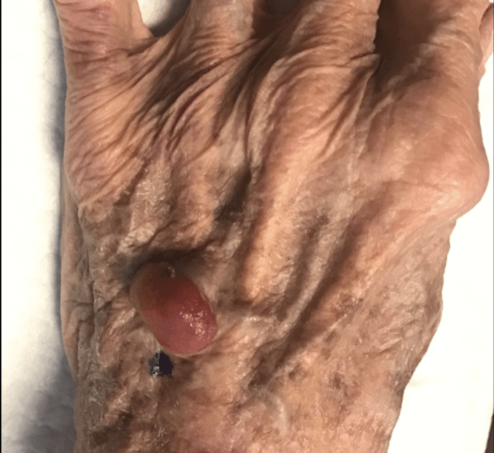
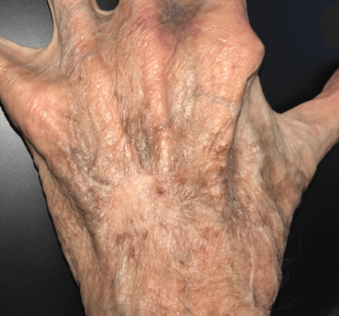
Melanoma Before and After*
See More ResultsWho is susceptible to being diagnosed with melanoma?
Anyone can be susceptible to being diagnosed with melanoma, though persons with a family history or personal history of melanoma, history of prolonged UV light exposure, having many moles or skin lesions, or those with fair skin do have a higher tendency of developing melanoma. Even if a person has any or all of these risk factors, it does not mean they will automatically develop melanoma at some point in their lives. Regular skin checks or annual/semi-annual dermatological skin checks can help with the clinical evaluation of moles, other skin lesions, and can lead to a more early diagnosis of skin cancers.
Where is Melanoma found?
Melanoma can be found anywhere on the body, but most often it is located on the chest and back of men and the legs of women. However, melanoma can be found in any area of the body, including the eyes, mouth, palms of the hands, and soles of the feet. These findings are rare, but can still occur. Again, melanoma usually presents as a darkened skin pigment that may or may not grow, change or bleed.
Melanoma Faqs
How serious is a melanoma diagnosis?
When a patient is diagnosed with melanoma, it will be diagnosed in a stage. The earliest diagnosis of melanoma is considered melanoma in situ or at Stage 0. The “in situ” diagnosis confirms that the melanoma is “in the cell” or confined within the epidermis and has not spread anywhere else in the body. If the melanoma is diagnosed after this stage, the next progressions of diagnoses are Stages I-IV. Within each of these stages are clinical recommendations for treatments and depth of invasion. For further reading and understanding of melanoma staging and diagnosis, please visit the American Cancer Society through this link. Furthermore, if you or a loved one have been diagnosed with melanoma of any stage, please seek medical treatment as this type of cancer can become metastatic and deadly.
What treatment options are available?
Developing a treatment plan is dependent upon the stage of melanoma diagnosis. If caught early enough, surgical intervention can be sufficient. If the melanoma can be surgically removed, our patients can move forward with the excision of the melanoma in our office on an outpatient basis and under only local anesthesia. Our office specializes in the removal of melanoma, and we can send specimens to a dermatopathologist or a doctor who specializes in dermatological specimens. After the specimen(s) are excised, Dr. Mountcastle will either leave the area open until we have confirmed negative margins (no remaining melanoma) from the pathology lab (which usually takes 3-5 business days) or place sutures into the area in which he excised and wait for the pathology results. If necessary, it is possible for Dr. Mountcastle to proceed with a re-excision if there are still positive margins (remaining melanoma). However, each patient is unique, as is each melanoma diagnosis. Depending on the location and depth of invasion of the melanoma, it may be necessary for Dr. Mountcastle to perform a skin grafting of the area if there is not enough epidermis to close the wound. Other treatment options for more progressively staged melanoma include, but are not limited to: sentinel node biopsies, full thickness skin grafting, and radiation therapy, all of which are performed in a hospital setting. If the melanoma has progressed to these stages, additional clinical staff such as a medical oncologist and a radiation oncologist may be necessary for accurate and efficient treatment planning. For further readings on melanoma treatments, please visit the American Cancer Society through this link.
Financing Available
Financing InfoRequest An Appointment
Contact Us